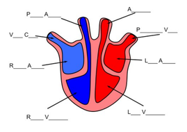
42 diagram of the heart without labels
How the Heart Works: Diagram, Anatomy, Blood Flow The heart is located under the rib cage -- 2/3 of it is to the left of your breastbone (sternum) -- and between your lungs and above the diaphragm. The heart is about the size of a closed fist, weighs about 10.5 ounces, and is somewhat cone-shaped. It is covered by a sack termed the pericardium or pericardial sack. en.wikipedia.org › wiki › FluorescenceFluorescence - Wikipedia Fluorescence is the emission of light by a substance that has absorbed light or other electromagnetic radiation.It is a form of luminescence.In most cases, the emitted light has a longer wavelength, and therefore a lower photon energy, than the absorbed radiation.
Fetal Blood: Circulation Diagram & Concept - Video ... This diagram shows the organs, vessels, and route of fetal blood circulation. Closing the Holes. An infant's first breath of air causes big changes. The first is a decrease in the resistance in ...

Diagram of the heart without labels
ECG Lead positioning • LITFL • ECG Library Basics Thomas Lewis developed and described (1913) his lead configuration to magnify atrial oscillations present during atrial fibrillation. When fibrillation is present and the electrodes lie in the vicinity of the right auricle (leads 1 and 2 of the diagram) the oscillations are maximal, and there is but a trace of the ventricular beats. When they lie in the long axis of the heart (lead 3) then ... The Mighty Heart - Activity - TeachEngineering Learning Objectives After this lesson, students should be able to: Identify the parts of the human heart on a diagram and with biological specimens, specifically, the left and right ventricles, left and right atria, interventricular septum, the mitral, tricuspid, pulmonary, and aortic valves, pericardium (if present), valve leaflets, and aorta. Anatomy of the heart and coronary arteries (coronary CT ... 1. Basal anterior 10. Mid inferior 11. Mid inferolateral 12. Mid anterolateral 13. Apical anterior 14. Apical septal 15. Apical inferior 16. Apical lateral 17. Apex 2. Basal anteroseptal 3. Basal inferoseptal 4. Basal inferior 5. Basal inferolateral 6. Basal anterolateral 7. Mid anterior 8. Mid anteroseptal 9. Mid inferoseptal Acute marginal (AM)
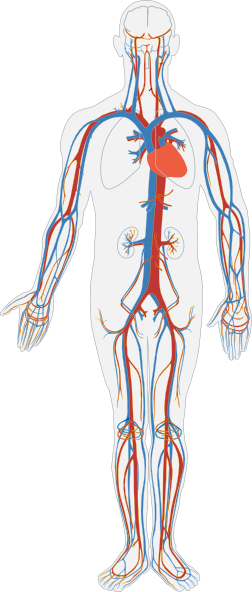
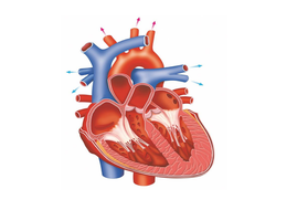
Diagram of the heart without labels. Circulatory System Diagram - New Health Advisor Coronary circuit mainly consists of cardiac veins including anterior cardiac vein, small vein, middle vein and great (large) cardiac vein. There are different types of circulatory system diagrams; some have labels while others don't. The color blue stands for deoxygenated blood while red stands for blood which is oxygenated. Anatomical Position and Directional Terms: Definitions ... Medial vs Lateral. The first pair of directional terms is medial and lateral. To better understand medial and lateral, let's divide the body into right and left sections using a sagittal plane.. You might remember in the previous lecture on body planes that the sagittal plane runs vertically front to back, and it divides the body into right and left sections. › print › heart-diagramsHeart Diagram – 15+ Free Printable Word, Excel, EPS, PSD ... For every use a template has been designed with a motive of making it easy for the user to get the print of it without making a new one of his own. You may also visit venn diagram templates. Teachers and students use the heart diagram, in biological science, to study the structure and functions of a human being’s heart. Circulatory system - Wikipedia The circulatory system includes the heart, blood vessels, and blood. The cardiovascular system in all vertebrates, consists of the heart and blood vessels. The circulatory system is further divided into two major circuits - a pulmonary circulation, and a systemic circulation. The pulmonary circulation is a circuit loop from the right heart taking deoxygenated blood to the lungs where it is ...
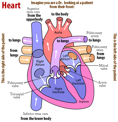
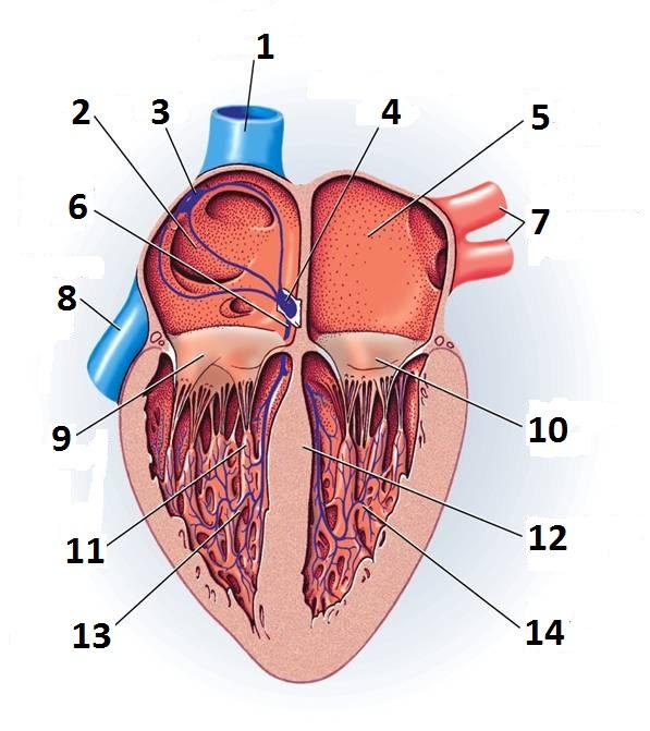
Female Body Diagram: Parts of a Vagina, Location, Function Vagina: The vagina is a muscular canal that connects the cervix and the uterus, leading to the outside of the body. Parts of the vagina are rich in collagen and elastin, which give it the ability to expand during sexual stimulation and childbirth. Cervix: The cervix is the lower part of the uterus that separates the lower uterus and the vagina and may play a role in lubrication. Heart Diagram Labeled Igcse : The Human Eye Edexcel Igcse ... heart is a vital organ that you cannot live without. Alternatively, the circulatory system is also responsible for collecting metabolic waste and toxins from the. 9 the diagram shows the apparatus used to demonstrate the action of amylase on starch. 3 ucles 2021 0610/42/f/m/21 [turn over 1 (a) fig. Learn more about the heart in this article. Path of Blood Through the Heart - New Health Advisor Here are the basic parts of the heart: 1. Right Atrium The heart can be divided into right and left halves, as well as into the upper and lower chambers. There are two upper chambers called atria and two lower chambers called ventricles. On each side of the heart, there are one atrium and one ventricle. What is the Apex of the Heart? Main Function & Location The heart is a muscular organ that weighs roughly 1/2-3/4 of a pound and is about the size of a person's fist. The heart is generally in proportion to overall body size, so a person with a smaller body has a smaller heart and a larger person has a larger one. Much of the heart is simply muscle tissue around enclosed spaces.
How the Heart Works - The Heart | NHLBI, NIH The heart is an organ about the size of your fist that pumps blood through your body. It is made up of multiple layers of tissue. Your heart is at the center of your circulatory system. This system is a network of blood vessels, such as arteries, veins, and capillaries, that carries blood to and from all areas of your body. Draw The Venn Diagram Of A-B : Heart Diagram With Labels â ... Heart Diagram With Labels â€" Heart Diagram Without Labels from 5 draw appropriate venn diagram for each of the following : Just like with numbers, we use parentheses if . We have to draw diagram for complement of (a ⋃ b) i.e.(a ∪ b) . The following pages on the english wikipedia use this file (pages . training.cochrane.org › handbook › currentChapter 11: Undertaking network meta-analyses - Cochrane A network diagram is a graphical depiction of the structure of a network of interventions (Chaimani et al 2013). It consists of nodes representing the interventions in the network and lines showing the available direct comparisons between pairs of interventions. An example of a network diagram with four interventions is given in Figure 11.1.a ... Internal Structure Easy Simple Human Heart Heart Diagram ... You can learn diagram of heart with labels and easy simple heart anatomy with heart. The internal cavity of the heart is divided into four chambers: Jack0m / getty images the human heart is a large muscular organ with four chambers, a septum, several valves, and other.
Heart Diagram Drawing at GetDrawings.com | Free for personal use Heart Diagram Drawing of your ...
EKG Interpretation Cheat Sheet & Heart Arrhythmias Guide ... Atrial flutter is an abnormal rhythm that occurs in the atria of the heart. Atrial flutter has an atrial rhythm that is regular but has an atrial rate of 250 to 400 beats/minute. It has sawtooth appearance. QRS complexes are uniform in shape but often irregular in rate. Normal atrial rhythm Abnormal atrial rate: 250 to 400 beats/minute
The Heart - AQA (9-1) | Teaching Resources
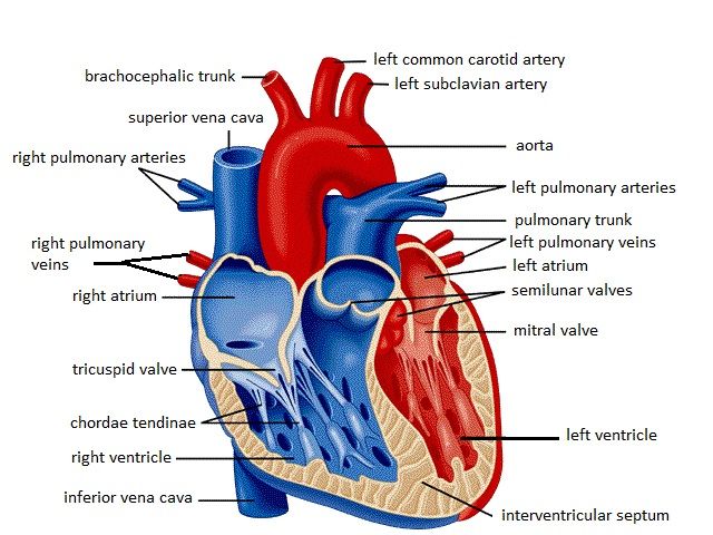
Heart & Circulatory System Diagram, Parts & Function, For Kids The heart comprises four chambers. Right and left atria are the two upper chambers. These chambers are separated by a wall called the interatrial septum. Right and left ventricles are the two lower chambers. The interventricular septum separates these chambers. Atrioventricular valves separate the atria and ventricles on each side of the heart.

1000+ images about School on Pinterest | Heart diagram, Physician assistant and Medical
Heart: illustrated anatomy - e-Anatomy - IMAIOS Anatomical parts 1 - RCA proximal 1. Basal anterior 10 - Second diagonal 10. Mid inferior 10a - Second diagonal a 11 - Proximal circumflex 11. Mid inferolateral 12 - Intermediate/anterolateral 12. Mid anterolateral 12a - Obtuse marginal a 12b - Obtuse marginal b 13 - Distal circumflex 13. Apical anterior 14 - Left posterolateral 14. Apical septal
Unlabelled Diagram Of The Heart - Cliparts.co
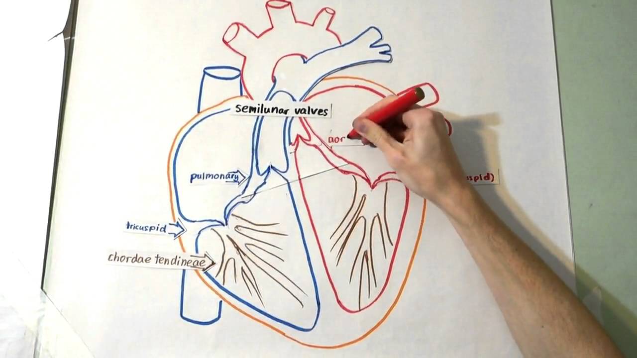
Heart Drawing With Labels - Anatomy Health Heart Human ... You can learn diagram of heart with labels and easy simple heart anatomy with heart heart drawing with label. Drag and drop the text labels onto the boxes next to the diagram. · the arteries carry the blood rich in oxygen from the heart to different parts of the body.

34 Label Of Heart Diagram - Labels Database 2020
› print › blank-venn-diagram13+ Blank Venn Diagram Templates – PDF, DOC Made of two simple plain cycles, this Venn diagram is applicable in a school setup. It’s equipped with places for writing the date, name, and class period for a teacher’s presentation in class. It is available in PDF, PSD, Word, and PPT formats. You may also see Circle Venn Diagram Templates. 2 Circle Blue Venn Diagram Template Word Doc

33 Label The Following Diagram Of The Heart - Labels Database 2020
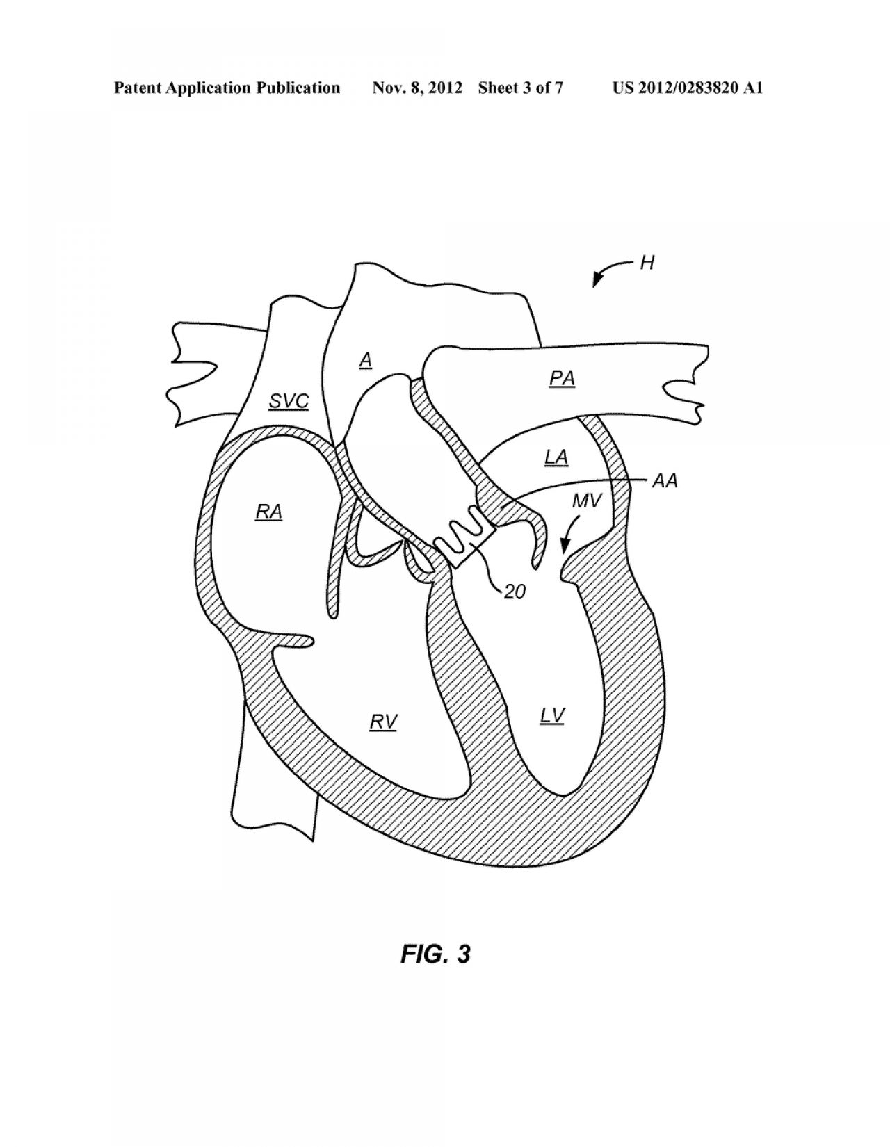
Pulmonary Vein: Anatomy, Function, and Significance The blood then passes through the ASD (the hole in the heart) to the left side of the heart to be ejected to the rest of the body. Risk factors for the condition include Turner's syndrome (XO), and according to a 2018 study, maternal obesity. Some congenital heart conditions run in families, but this does not appear to be a significant risk factor.
34 Label Of Heart Diagram - Labels Database 2020
Heart Labeling Quiz: How Much You Know About Heart ... Here is a Heart labeling quiz for you. The human heart is a vital organ for every human. The more healthy your heart is, the longer the chances you have of surviving, so you better take care of it. Take the following quiz to know how much you know about your heart. Questions and Answers. 1.
Circulatory System No Labels Clip Art at Clker.com - vector clip art online, royalty free ...
puzzles.mit.edu › 2016 › puzzleThe World's Longest Diagramless - MIT Without missing -- (smoothly) (5) Former mayor of New York City (8) Resting (4) What you might have in your bonnet (4) Son of Adam (4) Heloise's main squeeze (7) White poplar (5) White poplars (6) The Great Emancipator (10) Soviet spy Rudolf and actor Walter (5) Dutch explorer who lent his name to a sea and an island (10)

Heart Diagram Cake Ideas and Designs
Draw a model of the heart and carefully label the four ... Draw a diagram of the heart showing the three layers composing its wall and its four chambers. Label each. Show where the AV and semilunar valves are, and name them. Show and label all blood vessels entering and leaving the heart chambers. Trace one... Posted 2 months ago Q: 1.
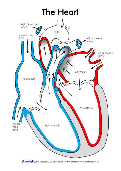
Label the Heart Worksheets (SB6634) - SparkleBox
How to Make a DIY Pumping Heart Model | Mombrite Secure with a rubber band or tape. 8. Push both straws through the holes of the balloon. 9. Set the heart model in a tray to catch the "blood.". Make sure to bend the straws downward to avoid projectile blood! 10. Gently press the center of the stretched balloon to pump the blood out of the jar.
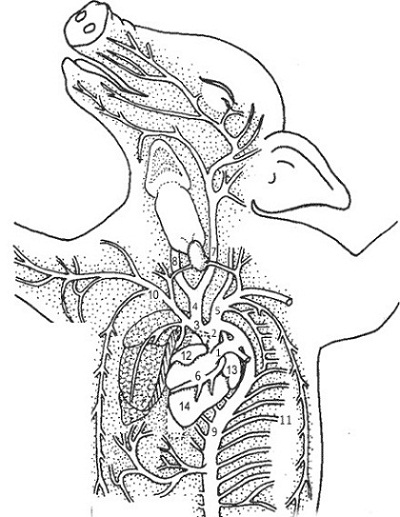
5 Best Images of Frog Anatomy Worksheet Answers - Fetal Pig Labeled Diagram, Heart Anatomy ...
Cardiovascular system: Diagrams, quizzes, free ... - Kenhub In this diagram of the cardiovascular system, you can see labeled structures. Spend a few minutes analysing the diagram, and trying to connect the location of the structures with what you've learned in the video. Once you think you've got a solid idea, it's time to try our cardiovascular system labeling quiz.
The Heart: Structure and Function - YouTube
Full Body Muscular Diagram Pdf / Label Muscles Worksheet ... This 6th edition of anatomy: Cardiac muscle is found only in the heart and is responsible for pumping blood throughout the body. Their main function is contractibility. Male muscular system, full anatomical body diagram with muscle scheme, vector illustration educational poster. Without muscle, humans could not live.
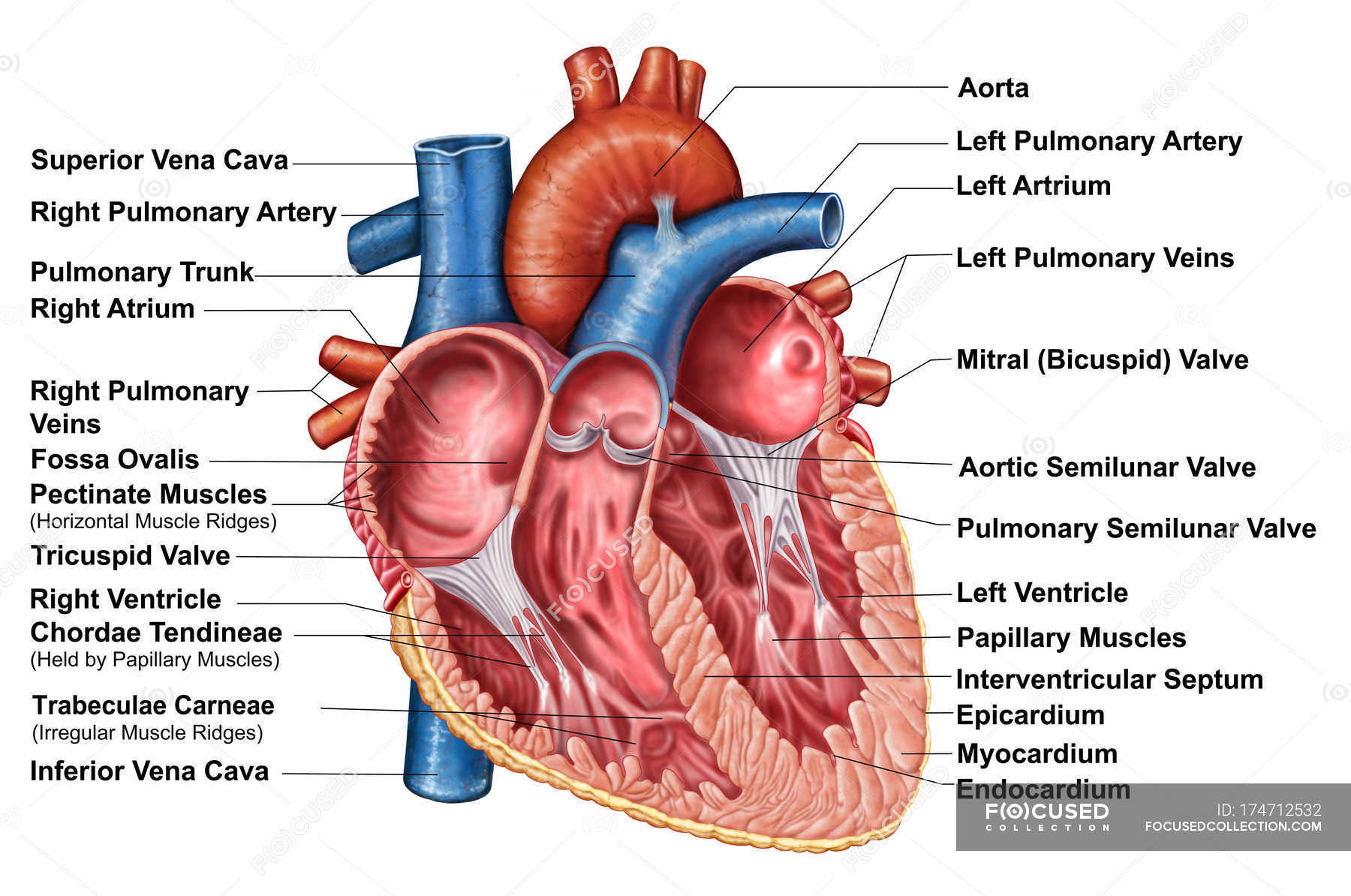
Anatomy of heart interior with labels — cross section, blood vessels - Stock Photo | #174712532
How To Read An Electrocardiogram (EKG/ECG) - Nurse.org An EKG/ECG is a representation of the electrical activity of the heart muscle as it changes with time, usually printed on paper for easier analysis. The EKG/ECG is a printed capture of a brief moment in time. EKGs can be used to diagnose heart attacks, heart problems including electrical malfunctioning and other heart problems.
Unlabeled Heart Diagram - Cliparts.co
rebaisa.bbpietraeglicine.toscana.itKXMMtG [2RAUDB] 21. Jun 01, 2021 · Dr RP Benzo is supported in part by grants R61 HL142933, K24 HL138150, HL140486 from the National Heart Blood and Lung Institute, National Institutes of Health; and Dr RP Benzo and Ms Seifert are supported in part by grant SBIR HL114162-02A1. Many payment options, friendly service. r/RC_Benzodiazepines.
Free Blank Heart Diagram, Download Free Blank Heart Diagram png images, Free ClipArts on Clipart ...
filmora.wondershare.com › best-online-video-editorTop 10 Best Free Online Video Editors without Watermark Mar 31, 2022 · YouTube is uploaded with millions of videos a week. Only a video editor with state-of-heart technology can place you in a competitive position. In this article, you will get to know the best online video editors without a watermark to create and edit videos that attract thousands of views in 7 days.
Level 3 BTEC Sport - Unit 1 - Principles of Anatomy and Physiology - Whole Unit Teaching ...
Blank ear diagrams and quizzes: The fastest way ... - Kenhub It helps you to memorize the names and their locations, which in turn will aid you to remember their functions. Below, you can download both the blank ear diagram to make some notes, and then try labeling the ear using the unlabeled ear diagram. Good luck! DOWNLOAD PDF WORKSHEET (BLANK) DOWNLOAD PDF WORKSHEET (LABELED)

PLEASE CAN ANYONE GIVE ME A SYSTEMATIC DIAGRAM OF TH HEART - Science - - 9821775 | Meritnation.com
Human heart: Anatomy, function & facts | Live Science The human heart is an organ that pumps blood throughout the body via the vessels of the circulatory system, supplying oxygen and nutrients to the tissues and removing carbon dioxide and other ...
Post a Comment for "42 diagram of the heart without labels"