40 diagram of the brain without labels
A Neurosurgeon's Overview the Brain's Anatomy The brainstem is the lower extension of the brain, located in front of the cerebellum and connected to the spinal cord. It consists of three structures: the midbrain, pons and medulla oblongata. It serves as a relay station, passing messages back and forth between various parts of the body and the cerebral cortex. Parts of the brain: Learn with diagrams and quizzes - Kenhub Labeled brain diagram First up, have a look at the labeled brain structures on the image below. Try to memorize the name and location of each structure, then proceed to test yourself with the blank brain diagram provided below. Labeled diagram showing the main parts of the brain Blank brain diagram (free download!)
How to make a Brain Model - Human Body Science for Kids Brain model with labels The outer part of the brain is called the Cerebrum, which is divided into two hemispheres, by a central fissure. Each Hemisphere is split into the 4 lobes, as shown above. Under the Cerebrum are the Cerebellum and the Brain Stem. The brain can be divided up into six main areas: The frontal lobe

Diagram of the brain without labels
Parts of the Brain Activity for Kids, Brain Diagram, and Worksheets for ... Learn about the Parts of the Brain for Kids with this fun brain activity for kids and handy brain worksheets! Children will learn about the functions of the brain for kids as part of our human body lesson. Use this brain project with elementary age students in first grade, 2nd grade, 3rd grade, 4th grade, 5th grade, and 6th garde students. Human Brain Diagram - Labeled, Unlabled, and Blank May 25, 2016 - Click here to download a free human brain diagram. Learn the parts of the human brain with these convenient printables for students and teachers. human body anatomy with labels Stock Photos and Images Set of brain and heart, kidney, colon logo or insignia, emblems, labels and badge. Medical icon design vector Abstract creative concept vector line draw background for web, mobile app, illustration template design, business infographic, page, brochure, banner, presentation, poster, cover, booklet, document.
Diagram of the brain without labels. Diagram Of Brain with their Labelings and Detailed Explanation The midbrain is the smallest region of the brain, found at the centre of the brain, between cerebral cortex and hindbrain. It comprises tectum, cerebral peduncle, tegmentum, cerebral aqueduct, substantia nigra, several nuclei and fasciculi. The midbrain is responsible for hearing, vision, sleep cycle, temperature regulation, alertness, etc. Human Nervous System - Diagram - How It Works | Live Science It consists of the brain, spinal cord and the retinas of the eyes. The Peripheral Nervous System consists of sensory neurons, ganglia (clusters of neurons) and nerves that connect the central... Label Brain Diagram Printout - EnchantedLearning.com Label the Brain Anatomy Diagram The Brain Read the definitions below, then label the brain anatomy diagram. Cerebellum - the part of the brain below the back of the cerebrum. It regulates balance, posture, movement, and muscle coordination. Corpus Callosum - a large bundle of nerve fibers that connect the left and right cerebral hemispheres. Brain Label - The Biology Corner This work is licensed under a Creative Commons Attribution-NonCommercial-ShareAlike 4.0 International License. Answers: A = parietal labe | B = gyrus of the cerebrum | C = corpus callosum | D = frontal lobe E = thalamus | F = hypothalamus | G = pituitary gland | H = midbrain
Neuron Diagram Unlabeled neuron, (1). axon, cell body, dendrites, nucleus, terminal. Unlabeled diagram of a motor neuron (try labeling: axon, dendrite, cell body, myelin, nodes of Ranvier, motor end plate).Read the definitions, then label the neuron diagram below. axon - the long extension of a neuron that carries nerve impulses away from the body of the cell. Picture of the Brain - WebMD • The cortex is the outermost layer of brain cells. Thinking and voluntary movements begin in the cortex. • The brain stem is between the spinal cord and the rest of the brain. Basic functions like... Heart Diagram No Labels Teaching Resources | Teachers Pay Teachers DrM. $4.00. PDF. Product DescriptionThis resource includes all the information you will need to help your students label the basic structures of the heart. It also includes two versions of the oversized diagram (labeled and unlabeled), as well as basic notes and a Exploration Guide to accompany the diagrams. Brain: Atlas of human anatomy with MRI - e-Anatomy - IMAIOS Anatomy of the brain (MRI) - cross-sectional atlas of human anatomy. The module on the anatomy of the brain based on MRI with axial slices was redesigned, having received multiple requests from users for coronal and sagittal slices. The elaboration of this new module, its labeling of more than 524 structures on 379 MRI images in three different ...
Human Brain Diagrams and Detailed Information - Innerbody The brainstem is made of three regions: the medulla oblongata, the pons, and the midbrain. A net-like structure of mixed gray and white matter known as the reticular formation is found in all three regions of the brainstem. The reticular formation controls muscle tone in the body and acts as the switch between consciousness and sleep in the brain. Diagram of the Brain and its Functions - Bodytomy Given below is a labeled diagram showing the brain stem and its related structures. Brain Stem and Structures Cerebellum The word 'cerebellum' literally means little brain. It is the second largest part of the brain, and is located at the back, below the occipital lobe, beneath the cerebrum and behind the brain stem. Anatomical diagrams of the brain - e-Anatomy - IMAIOS These original illustrations and diagrams of the brain were created from 3D medical imaging reconstructions and then redrawn and colored using Adobe Illustrator. These anatomical charts include the main diagrams necessary for medical students, nursing students, residents, practitioners, anatomists to study the anatomy of the brain, to ... Nervous System - Label the Brain - TheInspiredInstructor.com Nervous System - Label the Brain Nervous System - Brain Name: Choose the correct names for the parts of the brain. ( 1) (2) (3) (4) (5) (6) (7) (8) ( 9) This brain part controls thinking. (10) This brain part controls balance, movement, and coordination. (11) This brain part controls involuntary actions such as breathing, heartbeats, and digestion.
Where It's AT: Mrs. DiChiara's Technology Blog: Dyslexia and the Brain
75,682 Brain Anatomy Stock Photos and Images - 123RF Brain Anatomy Stock Photos And Images. 75,682 matches. Page of 757. Brain lobes vector illustration. Human brain infographic vector. Brain lobes functions. Serotonin pathway. Humans brain with serotonin pathways. psychiatric and neurological disorders. 3D render of a medical image showing male figure with brain tumour.
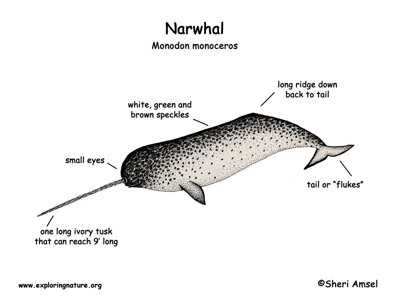
Narwhal
Structure, Diagram, Parts Of Human Brain - BYJUS The brain diagram given below highlights the different lobes of the human brain. Where is the Brain located? The brain is enclosed within the skull, which provides frontal, lateral and dorsal protection. The skull consists of 22 bones, 14 of which form the facial bones and the remaining 8 form the cranial bones.
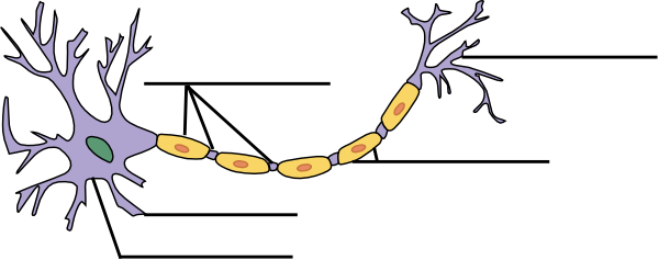
Label The Neuron Clip Art at Clker.com - vector clip art online, royalty free & public domain
The Human Brain (Diagram) (Worksheet) | Therapist Aid The Human Brain Diagram is a versatile tool for psychoeducation. The diagram separates the brain into six major parts, and provides a brief description of the functions carried out by each section. Discussion of the brain, and how it works, can be a powerful way to explore many topics. Explaining the physical changes behind mental illness can ...

Lacrosse Stadium as an Animal Cell
Labeled Diagrams of the Human Brain You'll Want to Copy Now The average dimension of the adult human brain is 5.5 inches in width and 6.5 inches in length. The height of the human brain is about 3.6 inches and it weighs about 4 to 5 lbs at birth and 3 lbs in adults. The total surface area of the cerebral cortex is about 2,500 cm2 and when stretched, it will cover the area of a night table.
Post a Comment for "40 diagram of the brain without labels"