42 human eye with labels
Eye Diagram - Differentiated Worksheets and EASEL Activities - Pinterest Description Use these simple eye diagrams to help students learn about the human eye. Three differentiated worksheets are included: 1. Write the words using a word bank 2. Cut and paste the words 3. Write the words without a word bank Labels include: eyebrow, eyelid, eyelashes, pupil, iris, and sclera. Labelled Diagram of Human Eye, Explanation and Function - VEDANTU The human eye is a part of the sensory nervous system. Labeled Diagram of Human Eye The eyes of all mammals consist of a non-image-forming photosensitive ganglion within the retina which receives light, adjusts the dimensions of the pupil, regulates the availability of melatonin hormones, and also entertains the body clock.
Human Eye Diagram, How The Eye Work -15 Amazing Facts of Eye Fun Facts About Human Eye For Kids FACT 1 Iris scanning is more secure than fingerprints because our iris has 256 unique characteristics and the fingerprint has just 40. FACT 2 Newborn babies don't produce tears. They only make crying sounds, but no tears come out of their crying eyes.

Human eye with labels
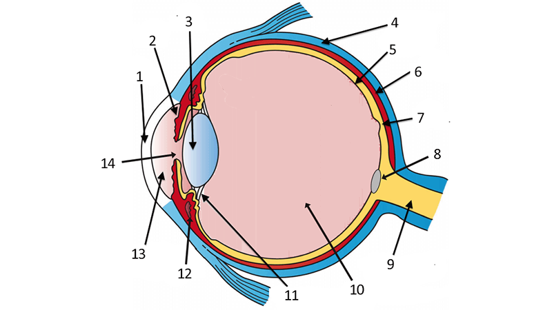
Structure and Function of the Human Eye - ThoughtCo The main parts of the human eye are the cornea, iris, pupil, aqueous humor, lens, vitreous humor, retina, and optic nerve. Light enters the eye by passing through the transparent cornea and aqueous humor. The iris controls the size of the pupil, which is the opening that allows light to enter the lens. Light is focused by the lens and goes ... The Eyes (Human Anatomy): Diagram, Optic Nerve, Iris, Cornea ... - WebMD The front part (what you see in the mirror) includes: Iris: the colored part. Cornea: a clear dome over the iris. Pupil: the black circular opening in the iris that lets light in. Sclera: the ... The Human Eye | Boundless Physics | | Course Hero The Human Eye The human eye is an organ that reacts with light and allows light perception, color vision and depth perception. Learning Objectives Identify parts of human eye and their functions Key Takeaways Key Points The eye is made up of a number of parts, including the iris, pupil, cornea, and retina.
Human eye with labels. Labeled Eye Diagram | Eye anatomy diagram, Eye anatomy ... - Pinterest This Article is the detailed account of all the major organs that are categorized under the nine regions in the abdominal cavity 1) Stomach 2) Intestines a) Small Intestine Duodenum Jejunum Ileum b) Large Intestine Ceacum Colon (Ascending, Transverse and Descending) Rectum Anal Canal 3) Liver 4) Gall bladder 5) Pancreas 6) Spleen 7) Kidneys […] S Human eye - Wikipedia The human eye is a sensory organ, part of the sensory nervous system, that reacts to visible light and allows us to use visual information for various purposes including seeing things, keeping our balance, and maintaining circadian rhythm.. The eye can be considered as a living optical device.It is approximately spherical in shape, with its outer layers, such as the outermost, white part of ... Labelling the eye — Science Learning Hub Labelling the eye Add to collection The human eye contains structures that allow it to perceive light, movement and colour differences. In this activity, students use online or paper resources to identity and label the main parts of the human eye. By the end of this activity, students should be able to: identify the main parts of the human eye File:Diagram of human eye without labels.svg - Wikimedia Size of this PNG preview of this SVG file: 410 × 430 pixels. Other resolutions: 229 × 240 pixels | 458 × 480 pixels | 732 × 768 pixels | 976 × 1,024 pixels | 1,953 × 2,048 pixels. Original file (SVG file, nominally 410 × 430 pixels, file size: 277 KB) File information. Structured data.
human eye | Definition, Anatomy, Diagram, Function, & Facts human eye, in humans, specialized sense organ capable of receiving visual images, which are then carried to the brain. The eye is protected from mechanical injury by being enclosed in a socket, or orbit, which is made up of portions of several of the bones of the skull to form a four-sided pyramid, the apex of which points back into the head. Thus, the floor of the orbit is made up of parts of ... How to draw the Human Eye - Labeled Science Diagrams - YouTube Download a free printable outline of this video and draw along with us: you for watching. Please subsc... Eye Anatomy: 16 Parts of the Eye & Their Functions Apr 05, 2022 · The following are parts of the human eyes and their functions: 1. Conjunctiva. The conjunctiva is the membrane covering the sclera (white portion of your eye). The conjunctiva also covers the interior of your eyelids. Conjunctivitis, often known as pink eye, occurs when this thin membrane becomes inflamed or swollen. Eye Anatomy: Parts of the Eye and How We See Behind the anterior chamber is the eye's iris (the colored part of the eye) and the dark hole in the middle called the pupil. Muscles in the iris dilate (widen) or constrict (narrow) the pupil to control the amount of light reaching the back of the eye. Directly behind the pupil sits the lens. The lens focuses light toward the back of the eye.
Diagram human eye anatomy with label vector image Diagram of human eye anatomy with label illustration. Download a free preview or high-quality Adobe Illustrator (ai), EPS, PDF vectors and high-res JPEG and ... Eye anatomy: A closer look at the parts of the eye In a number of ways, the human eye works much like a digital camera: Light is focused primarily by the cornea — the clear front surface of the eye, which acts like a camera lens. The iris of the eye functions like the diaphragm of a camera, controlling the amount of light reaching the back of the eye by automatically adjusting the size of the ... Human Eye Anatomy Pictures, Images and Stock Photos The human eye is an organ that reacts to light and has several purposes. As a conscious sense organ, the mammalian eye allows vision. Engraving eyeball illustration on blue BG Engraving drawing human eyeball illustration isolated on blue turquoise background Human eye anatomy, right eye viewed from above Label Functions of Parts of the Human Eye Functions of the Parts of the Eye. Select the correct label for the function of each part of the eye. The image is taken from above the left eye. Click on the Score button to see how you did. Incorrect answers will be marked in red.
Discovering Something New -- ongoing learning: How the eye works
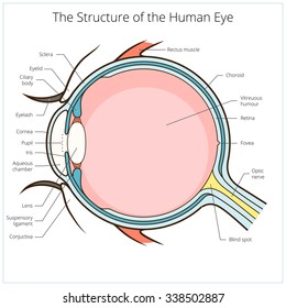
Anatomy of the Eye | Johns Hopkins Medicine Ciliary body. The part of the eye that produces aqueous humor. Cornea. The clear, dome-shaped surface that covers the front of the eye. Iris. The colored part of the eye. The iris is partly responsible for regulating the amount of light permitted to enter the eye. Lens (also called crystalline lens).

Human Anatomy Lab: Muscles of the Arm
Eye Diagram Quiz - ProProfs Try this amazing Eye Diagram Quiz quiz which has been attempted 4905 times by avid quiz takers. ... Label The Parts Of The Eye. People say that the eyes are the windows to a person's soul. ... How much did you get to understand about the human eye?... Questions: 8 | Attempts: 44712 | Last updated: Mar 22, 2022 . Sample Question. A is pointing ...

Moistcr1tikal Height, Weight, Age, Girlfriend, Facts, Biography - cornytube.com
Label Parts of the Human Eye Parts of the Eye Select the correct label for each part of the eye. The image is taken from above the left eye. Click on the Score button to see how you did. Incorrect answers will be marked in red.

Anatomy of the Eye
BYJUS BYJUS

Anatomy of the Human Eye - News-Medical Eyes are one of the most important organs of the body. ... Humans have binocular vision, meaning that both the eyes create a single combined image.
31 Label The Eye Quiz - Best Labeling Ideas
Labelling the eye — Science Learning Hub Feb 23, 2021 · The human eye has several structures that enable entering light energy to be converted to electrochemical energy. This stimulates the visual centres in the brain, giving us the sensation of seeing. In this interactive, you can label parts of the human eye. Use your mouse or finger to hover over a box to highlight the part to be named.

Human Eyeball Diagram - YouTube
Label the diagram of HUMAN EYE - Labelled diagram - Wordwall Glisează și fixează pinii în locul corect pe imagine.. SCLERA, CORNEA, PUPIL, LENS, IRIS, CHOROID, RETINA, OPTIC NERVE.
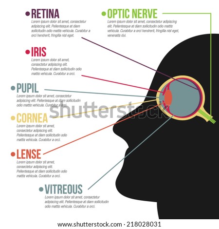
Basic Humans Eye Anatomy Vector Infographic Stock Vector (Royalty Free) 218028031 - Shutterstock
Labeled Eye Diagram | Science Trends The corneais the outermost portion of the eyeball. The responsibility of the cornea is to focus the light that enters our eyes. The cornea is transparent, and it covers the pupil, iris, and anterior chamber. The cornea itself is composed of five different layers, and the function of the outermost layer is to protect the eye from dirt and foreign ob...

33 Human Eye With Label - Labels Design Ideas 2020
Eye Diagram With Labels and detailed description - BYJUS A brief description of the eye along with a well-labelled diagram is given below for reference. Well-Labelled Diagram of Eye The anterior chamber of the eye is the space between the cornea and the iris and is filled with a lubricating fluid, aqueous humour. The vascular layer of the eye, known as the choroid contains the connective tissue.

6,782 Human eye diagram Images, Stock Photos & Vectors - Shutterstock Human eye diagram royalty-free images 6,782 human eye diagram stock photos, vectors, and illustrations are available royalty-free. See human eye diagram stock video clips Image type Orientation People Artists More Sort by Biology Healthcare and Medical Icons and Graphics Diseases, Viruses, and Disorders human eye anatomy eye 3d rendering retina

More Then 50 Best Tattoo Designs 2014 For Men ~ 8FACT
PDF Eye Anatomy Handout - National Eye Institute of light entering the eye. Lens: The lens is a clear part of the eye behind the iris that helps to focus light, or an image, on the retina. Macula: The macula is the small, sensitive area of the retina that gives central vision. It is located in the center of the retina. Optic nerve: The optic nerve is the largest sensory nerve of the eye.
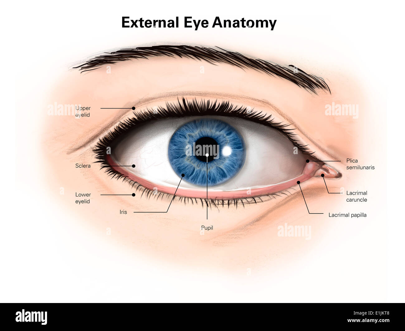
External anatomy of the human eye (with labels Stock Photo: 69866840 - Alamy
External anatomy of the human eye (with labels Stock Photo Download this stock image: External anatomy of the human eye (with labels). - E1JKT8 from Alamy's library of millions of high resolution stock photos, ...

World All Animals: Hippopotamuses New profile and Images
Labeling the Human Eye Quiz - PurposeGames.com About this Quiz. This is an online quiz called Labeling the Human Eye. There is a printable worksheet available for download here so you can take the quiz with pen and paper. This quiz has tags. Click on the tags below to find other quizzes on the same subject. Anatomy.
Easy Human Eye Diagram With Labels - Diagram Media
The Human Eye | Boundless Physics | | Course Hero The Human Eye The human eye is an organ that reacts with light and allows light perception, color vision and depth perception. Learning Objectives Identify parts of human eye and their functions Key Takeaways Key Points The eye is made up of a number of parts, including the iris, pupil, cornea, and retina.

Mickey's Artistry!: Cubism-My version of Houses at L'Estaque, Georges Braque
The Eyes (Human Anatomy): Diagram, Optic Nerve, Iris, Cornea ... - WebMD The front part (what you see in the mirror) includes: Iris: the colored part. Cornea: a clear dome over the iris. Pupil: the black circular opening in the iris that lets light in. Sclera: the ...

639 best images about Medical Terminology Help Learning on Pinterest
Structure and Function of the Human Eye - ThoughtCo The main parts of the human eye are the cornea, iris, pupil, aqueous humor, lens, vitreous humor, retina, and optic nerve. Light enters the eye by passing through the transparent cornea and aqueous humor. The iris controls the size of the pupil, which is the opening that allows light to enter the lens. Light is focused by the lens and goes ...

302 Found
Post a Comment for "42 human eye with labels"