44 simple microscope diagram with labels
Electron microscope - Wikipedia An electron microscope is a microscope that uses a beam of accelerated electrons as a source of illumination. As the wavelength of an electron can be up to 100,000 times shorter than that of visible light photons, electron microscopes have a higher resolving power than light microscopes and can reveal the structure of smaller objects. A scanning transmission electron microscope has achieved ... Labeling the Parts of the Microscope | Microscope World ... Labeling the Parts of the Microscope This activity has been designed for use in homes and schools. Each microscope layout (both blank and the version with answers) are available as PDF downloads. You can view a more in-depth review of each part of the microscope here. Download the Label the Parts of the Microscope PDF printable version here.
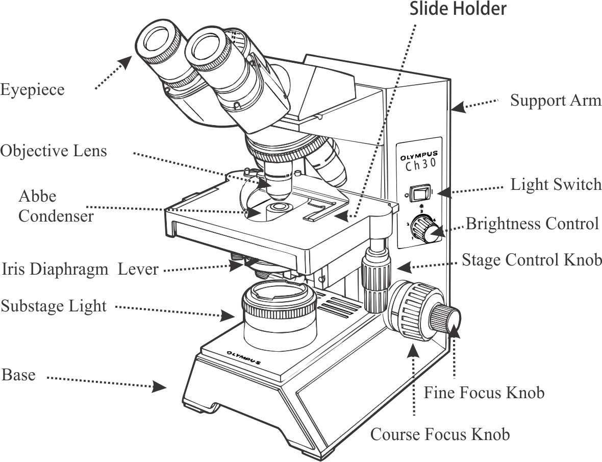
Parts of the Microscope with Labeling (also Free Printouts ... Parts of the Microscope with Labeling (also Free Printouts) A microscope is one of the invaluable tools in the laboratory setting. It is used to observe things that cannot be seen by the naked eye. Table of Contents 1. Eyepiece 2. Body tube/Head 3. Turret/Nose piece 4. Objective lenses 5. Knobs (fine and coarse) 6. Stage and stage clips 7. Aperture
Simple microscope diagram with labels
Labelled Diagram of Compound Microscope - Biology Discussion The below mentioned article provides a labelled diagram of compound microscope. Part # 1. The Stand: The stand is made up of a heavy foot which carries a curved inclinable limb or arm bearing the body tube. The foot is generally horse shoe-shaped structure (Fig. 2) which rests on table top or any other surface on which the microscope in kept. Compound Microscope- Definition, Labeled Diagram ... The optical microscope often referred to as the light microscope, is a type of microscope that uses visible light and a system of lenses to magnify images of small subjects. There are two basic types of optical microscopes: Simple microscopes. Compound microscopes. The term "compound" in compound microscopes refers to the microscope having ... A Study of the Microscope and its Functions With a Labeled ... sciencestruck.com A Study of the Microscope and its Functions With a Labeled Diagram To better understand the structure and function of a microscope, we need to take a look at the labeled microscope diagrams of the compound and electron microscope. These diagrams clearly explain the functioning of the microscopes along with their respective parts.
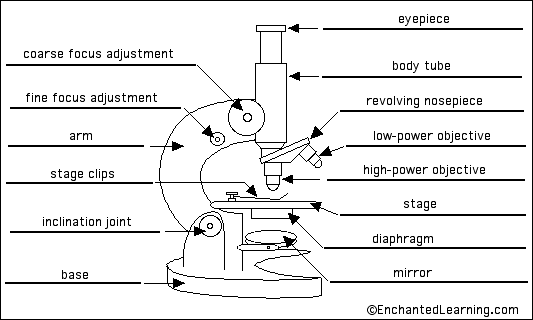
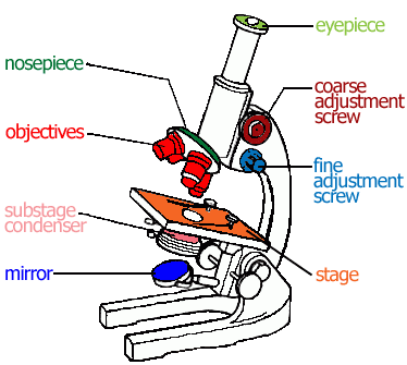
Simple microscope diagram with labels. Anatomy Chart - How to Make Medical Drawings and Illustrations Pathologic anatomy focuses on how diseases affect and change the human body. Histology studies microscopic anatomy such as tissues and cells visible only under a microscope. Anatomy charts serve two main purposes: education in the form of anatomy worksheets and presentation in the form of simple healthcare illustrations. Simple Columnar Epithelium Labeled Diagram Squamous means scale-like. simple squamous. Bodytomy provides a labeled diagram to help you understand the structure and Simple Columnar Epithelium: Labeled Diagram and Function. Epithelium is a tissue that lines the internal surface of the body, as well as the internal organs. Simple epithelium is one of the types of epithelium that is. Compound Microscope Parts - Labeled Diagram and their ... There are three major structural parts of a compound microscope. The head includes the upper part of the microscope, which houses the most critical optical components, and the eyepiece tube of the microscope. The base acts as the foundation of microscopes and houses the illuminator. The arm connects between the base and the head parts. PDF Parts of a Microscope Printables - Homeschool Creations Label the parts of the microscope. You can use the word bank below to fill in the blanks or cut and paste the words at the bottom. Microscope Created by Jolanthe @ HomeschoolCreations.net eyepiece head objective lenses arm focusing knob base illuminator stage stage clips nosepiece.
Microscope Poster - Diagram with Labels | Teach Starter A poster containing a diagram with labels showing the key parts of a microscope. In Science it is important that students know how to use a variety of tools when conducting scientific experiments and inquiry. This poster focuses on the microscope and highlights its key parts. There are two print options available for this poster: Microscope Labeling - The Biology Corner 1) Start with scanning (the shortest objective) and only use the COARSE knob . Once it is focused… 2) Switch to low power (medium) and only use the COARSE knob . You may need to recenter your slide. Once it is focused.. 3) Switch to high power (long objective). Fluorescence Resonance Energy Transfer (FRET) Microscopy Presented in Figure 3 is a Jablonski diagram illustrating the coupled transitions involved between the donor emission and acceptor absorbance in fluorescence resonance energy transfer. Absorption and emission transitions are represented by straight vertical arrows (green and red, respectively), while vibrational relaxation is indicated by wavy ... Compound Microscope Parts, Functions, and Labeled Diagram ... Compound Microscope Parts, Functions, and Labeled Diagram Parts of a Compound Microscope Each part of the compound microscope serves its own unique function, with each being important to the function of the scope as a whole.
Simple Microscope - Definition, Types, Working Principle ... A simple microscope consists of a convex lens of a short focal length. The below figure shows the ray diagram which subsequently forms the image of an object (or we can say a source of light). (Image will be Updated soon) F is the focal length of the lens. An object is placed between the focal length and the centre of the curvature. Parts of a microscope with functions and labeled diagram Structural parts of a microscope and their functions Figure created with biorender.com Figure: Diagram of parts of a microscope There are three structural parts of the microscope i.e. head, base, and arm. Head - This is also known as the body. It carries the optical parts in the upper part of the microscope. Base - It acts as microscopes support. Simple microscope - Fun Science Principle of Simple Microscope. A simple microscope works on the principle that when a tiny object is placed within its focus, a virtual, erect and magnified image of the object is formed at the least distance of distinct vision from the eye held close to the lens. Working of Simple Microscope. The ray diagram to show the working of simple ... Parts of a Simple Microscope - Labeled (with diagrams ... Parts of a Simple Microscope - Labeled (with diagrams) A simple microscope is a very first type of microscope ever created. It consists of simple parts and performs simple functions. Although there are now many advanced microscope types, some applications may still demand the use of a simple microscope.
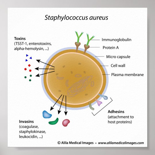
Staphylococcus aureus bacterium labeled diagram. poster | Zazzle.com
Microscope labeled diagram - SlideShare Microscope labeled diagram 1. The Microscope Image courtesy of: Microscopehelp.com Basic rules to using the microscope 1. You should always carry a microscope with two hands, one on the arm and the other under the base. 2. You should always start on the lowest power objective lens and should always leave the microscope on the low power lens ...

Microscope Unlabeled Diagram - Micropedia
Simple Microscope - Definition, Diagram, FAQs Simple Microscope Definition: A Simple Microscope meaning is used to see a magnified image of an object. Antonie Van Leeuwenhoek, a Dutchman, invented the first simple microscope, consisting of a single powerful magnetic lens that rotates to detect tiny freshwater insects. It is composed mainly of light microscopes.
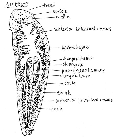
Flatworms
Looking at the Structure of Cells in the Microscope ... Many light-microscope techniques are available for observing cells. Cells that have been fixed and stained can be studied in a conventional light microscope, while antibodies coupled to fluorescent dyes can be used to locate specific molecules in cells in a fluorescence microscope. Living cells can be seen with phase-contrast, differential ...
The Microscope - General Revision for GCSE
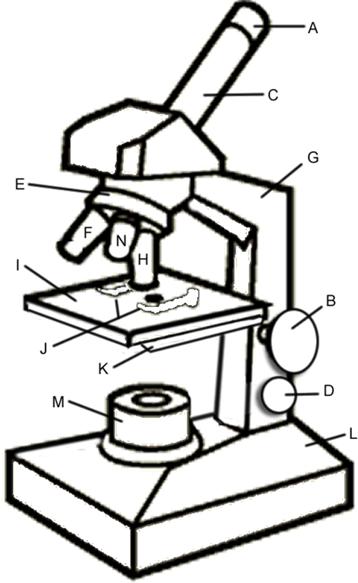
Label a Microscope - Storyboard That Create a poster that labels the parts of a microscope and includes descriptions of what each part does. Click "Start Assignment". Use a landscape poster layout (large or small). Search for a diagram of a microscope. Using arrows and textables label each part of the microscope and describe its function. Copy This Storyboard* More options
Compound Light Microscope Drawing | Free download on ClipArtMag
Botany Exam 1 Chs. 1, 2, 3, 4, 5, 6, 7, 12, and 16 Quizzes Mitosis, or the division of a mother cell's nucleus into two identical daughter nuclei, is typically divided into four phases. Match each of the labels to identify what events take place during each phase of mitosis. 1. Prophase 2. Metaphase 3. Anaphase 4. Telophase A) Chromosomes are aligned at the equator of the cell and the spindle is fully ...

the functions of a microscope | Diabetes Inc.
Interactive Bacteria Cell Model - CELLS alive Ribosomes: Ribosomes give the cytoplasm of bacteria a granular appearance in electron micrographs.Though smaller than the ribosomes in eukaryotic cells, these inclusions have a similar function in translating the genetic message in messenger RNA into the production of peptide sequences (proteins).
Light Microscope Diagram Labeled - Micropedia
Simple Microscope - Parts, Functions, Diagram and ... Simple Microscope - Parts, Functions, Diagram and Labelling A microscope is one of the commonly used equipment in a laboratory setting. A microscope is an optical instrument used to magnify an image of a tiny object; objects that are not visible to the human eyes. Table of Contents The common types of microscopes are: What is a Simple microscope?

Simple Microscope Labeled Diagram - Micropedia
Microscope Parts and Functions With Labeled Diagram and ... Microscope Parts and Functions With Labeled Diagram and Functions How does a Compound Microscope Work?. Before exploring microscope parts and functions, you should probably understand that the compound light microscope is more complicated than just a microscope with more than one lens.. First, the purpose of a microscope is to magnify a small object or to magnify the fine details of a larger ...

Pin on Diagramatically Speaking
Simple Microscope Definition, Magnification, Parts And Uses Aim: To make a simple microscope with the help of water. Apparatus Required A glass of water Fuse wire Object to view (newspaper works well due to its fine print) Procedure Make a loop of the fuse wire around 2 mm wide. Dip it in water so that a drop is made in the loop. Hold it near to your eye and take a close look at the object you have chosen.

Simple Cuboidal Epithelium Kidney Tubules In Cross Section 250x Shows Cuboidal Epithelium Lumens ...
Microscope Types (with labeled diagrams) and Functions Simple microscope labeled diagram Simple microscope functions It is used in industrial applications like: Watchmakers to assemble watches Cloth industry to count the number of threads or fibers in a cloth Jewelers to examine the finer parts of jewelry Miniature artists to examine and build their work Also used to inspect finer details on products

Microscope With Labels Clip Art at Clker.com - vector clip art online, royalty free & public domain
Label the Microscope Diagram | Download Scientific Diagram the antibiogram of e. coli was investigated in different generations using eight antibiotic discs such as chloramphenicol (ch), streptomycin (s), gentamycin (g), ciprofloxacin (ci),...

Simple Unlabelled Microscope Diagram - Micropedia
Label the microscope - Science Learning Hub All microscopes share features in common. In this interactive, you can label the different parts of a microscope. Use this with the Microscope parts activity to help students identify and label the main parts of a microscope and then describe their functions. Drag and drop the text labels onto the microscope diagram.

Cell Structure and Organisation
Labeling Microscope Worksheet | Teaching Resources A straightforward worksheet in which students are required to identify the parts of a basic microscope. Tes classic free licence. Reviews. 4.7 Something went wrong, please try again later. MACS0647-JD. a year ago. report. 5. Thanks. Very helpful. Empty reply does not make any sense for the end user ...

Chapter 1, Page 12 - HistologyOLM
Simple Microscope - Diagram (Parts labelled), Principle ... Parts of a Simple Microscope A simple microscope consists of Optical parts Mechanical parts Labeled Diagram of simple microscope parts Optical parts The optical parts of a simple microscope include Lens Mirror Eyepiece Lens A simple microscope uses biconvex lens to magnify the image of a specimen under focus.
Microscope Coloring - ClipArt Best - ClipArt Best
Microscope Drawing And Label at PaintingValley.com ... label microscope diagram compound parts light labeling functions microscopic blank labeled biology microscopy labelled beautiful Compound Microscope ... 496x600 35 0 Parts Of A Compound ... 500x469 27 0 Microscopic Drawing ... 1024x1024 21 4 Download The Diagram... 547x579 17 0 Microscope Labeling ... 270x350 17 0 Microscope Labeling ...
Post a Comment for "44 simple microscope diagram with labels"